- Definition
Community-acquired pneumonia is a common respiratory disease caused by a variety of bacteria, viruses, or even fungi. Radiographs are key to establishing the likely diagnosis and to start empiric treatment.
- Pathophysiology
The airway is not a sterile place. As the air enters and exits the lungs, countless microorganisms transit into the lungs. The pulmonary defenses keep the microbiome levels low and non-harmful. However, the organisms are able to multiply with weakened immune system or when a big reservoir is delivered (ie. aspiraiton of objects/food/water). When this happens, the macrophage responds by eating, then producing the inflammatory cytokines against the organisms. This response may spillover to body-wide war, as the cytokines travel to the bloodstream.
Some common typical organisms include: Streptococcus pneumoniae, Haemophilus influenzae, Pseudomonas aeruginosa, Staphylococcus aureus, respiratory viruses like COVID and influenza, RSV, parainfluenza, adenovirus.
Atypical organisms include: Mycobacter pneumoniae, Legionella pneumophila, Neisseria meningitidis, Mycobacterium tuberculosis, psittaci.
Risks:
- The winter months
- Older age
- Male
- Low socioeconomic population
- Symptoms & Signs
- Classic sudden fever, cough, and shortness of breath. Sputum may or may not be involved. Especially in the elderly population, mental status may also change, manifesting in a temporary state called delirium.
- Vital signs: tachycardia (faster heartbeat), tachypnea (faster breathing), hypoxemia, especially if the infection has spread systemically.


- Best Imaging Modality
-
- Clinical Suspicion
There are no certain rules for diagnosing pneumonia clinically; in fact, the diagnostic accuracy becomes severely limited without imaging. Nevertheless, if the patient is otherwise healthy and displays the following respiratory symptoms, a diagnosis could be made especially for patients who cannot receive an x-ray:
- fever
- cough
- consolidation detected on physical exam (whispering pectoriloquy test)
- wheeze
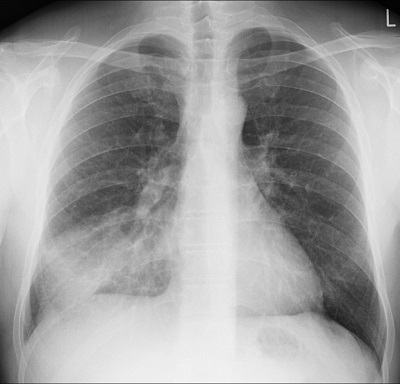
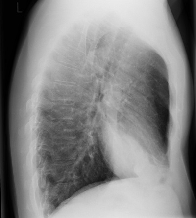
- Gold Standard: PA and Lateral Chest X-ray
Lobar consolidations (focal area of solidified tissue) can be found as a reaction to local infections. Interstitial infiltrates, which is the connective space between alveoli visible due to invasion of fluids, occur in pneumonia as an inflammatory response to infection. Cavitating lesions occur in some diseases such as tuberculosis, where gas-filled space develops with thick walls around it.
- Specificity
- Sensitivity: 100% @ 6 hours --> gradually decrease to 58% @ day 5.
- Pros: very quick, readily available, cheap
- Cons
The findings on CXR for pneumonia is not pathognomonic. Other differential diagnoses may include:
- Bronchitis
- Influenza, COVID
- Upper respiratory tract infection
- CT Chest
Taken when there is high clinical suspicion (ie. the symptoms are clearly pointing at a lung problem) but the chest x-ray does not show the disease. We could consider CT for patients with very weak immune system (that cannot mount a strong enough inflammatory response). The CT can also recognize the characteristic findings for certain organisms, helping narrow the diagnosis.
- Specificity
- Sensitivity: 83-98%
- Pros
- Cons
- Treatment
-
Treatment mainly focuses on antibiotics.
- Within 4 hours of arriving to the hospital, antibiotics should be given to optimize survival. Some options:
- Combination therapy: ceftriaxone, ampicillin, or ertapenem PLUS azithromycin or doxycycline.
- Single therapy: fluoroquinolone: levofloxacin / moxifloxacin / gemifloxacin
- Important coverage to consider: MRSA (methicillin resistant staphylococcus aureus) and pseudomonas.
References
- Torigian DA, Ramchandani P. Radiology Secrets Plus Fourth Edition. 2017. Elsevier.
- Klompas M. Clinical evaluation and diagnostic testing for community-acquired pneumonia in adults. UpToDate. 2021. (Accessed on August 2022)